by Luca Bell
Conventional T cells recognise peptide antigens presented by major histocompatibility complex (MHC) molecules, Human Leukocyte Antigen (HLA) molecules in humans. Unconventional T cell clones bind antigens restricted by MHC related protein 1 (MR1). Such cells, MAIT cells, are documented as recognising metabolic intermediates as antigens presented by MR1.
A group of researchers from around the globe reported a T cell receptor that recognised cancer cells and promoted cell lysis, while not recognising healthy cells. They also sought identify the mechanism of this selection. The T cells reported by the authors showed no difference in targeting engineered cancer cells that lacked peptide presenting molecules. This was similar to MAIT cells.
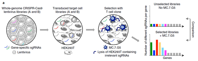
Peripheral blood mononuclear cells (PBMC) were isolated from blood by centrifuge and then cultured with lung carcinoma cells for two weeks. Primed PBMCs were cloned. These T cells are referred to as MC.7.G5. Embryonic kidney cells from the HEK293T cell line were transduced with a library of single guide RNAs (sgRNA) targeting all protein coding genes in the human genome. These sgRNAs direct the Cas9 nuclease to splice out the gene targeted by the sgRNA. This results in a library of HEK293T cells that each have a gene removed. These cells were incubated along with the MC.7.G5 T cells. HEK293T cells that survived were resistant to lysis by the MC.7.G5 T cells. The HEK293T cells were collected, and their DNA was isolated and sequenced using Illumina sequencing. These reads were compared to a control sample of HEK293T cells that were not incubated with MC.7.G5 T cells. sgRNAs that were enriched compared to the control indicated genes that were spliced out and resulted in increased survival. These genes are thus important for recognition by T cells.
The genes important for T cell recognition were found to encode proteins involved in promoter activation of MR1 and β2M, with which MR1 forms a dimeric antigen-presenting molecule known to activate MAIT cells and other T cells. MR1 being the primary antigen presenting molecule in the targeting of cancer cells by T cells was confirmed by a loss-of-function assay.
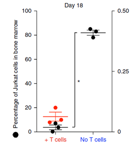
MC.7.G5 T cells were shown to decrease the Jurkat leukaemia cell burden in mice by an average of 95% compared to the control, after 18 days.
Benefits of MR1 as an antigen presenting molecule are that, unlike HLA molecules which are highly polymorphic and are present polymorphic peptide antigens, MR1 is monomorphic and presents non-peptide antigens.
In order to test the safety of MC.7.G5 T cells as a treatment, the T cells were incubated with healthy cells of different tissue types that had been stressed or infected with pathogens. MC.7.G5 T cells were found to be inert.
The take home message of this study is that MC.7.G5 T cells are an example of a potential cancer treatment that, unlike chemotherapy for example, does not target healthy cells as an adverse side effect and can be used to treat any human, regardless of their HLA genotype.
Reference
Crowther, M., Dolton, G., Legut, M., Caillaud, M., Lloyd, A., Attaf, M., Galloway, S., Rius, C., Farrell, C., Szomolay, B., Ager, A., Parker, A., Fuller, A., Donia, M., McCluskey, J., Rossjohn, J., Svane, I., Phillips, J. and Sewell, A., 2020. Genome-wide CRISPR–Cas9 screening reveals ubiquitous T cell cancer targeting via the monomorphic MHC class I-related protein MR1. Nature Immunology, 21(2), pp.178-185.

Leave a comment