A recent study by Miragaia and colleagues has shown that Treg cells in non-lymphoid tissue (NLT) such as skin and colon expresses a unique set of genes that are required for tissue adaptation which are very different from Treg cells in the lymphoid tissue (LT: specialized immunological tissues) i.e. lymph nodes and spleen. In this study they were able to identifying unique genes that could map out Treg cell trajectories as they transit from a specific tissue to another and thus were able to predict the fate of the cells.
To achieve this the authors performed single cell RNA-sequencing (scRNA-seq) of 3500 CD4+ T cell collected from skin, colon and associated lymph nodes using the droplet based 10x genomics technique as shown in Figure 1.
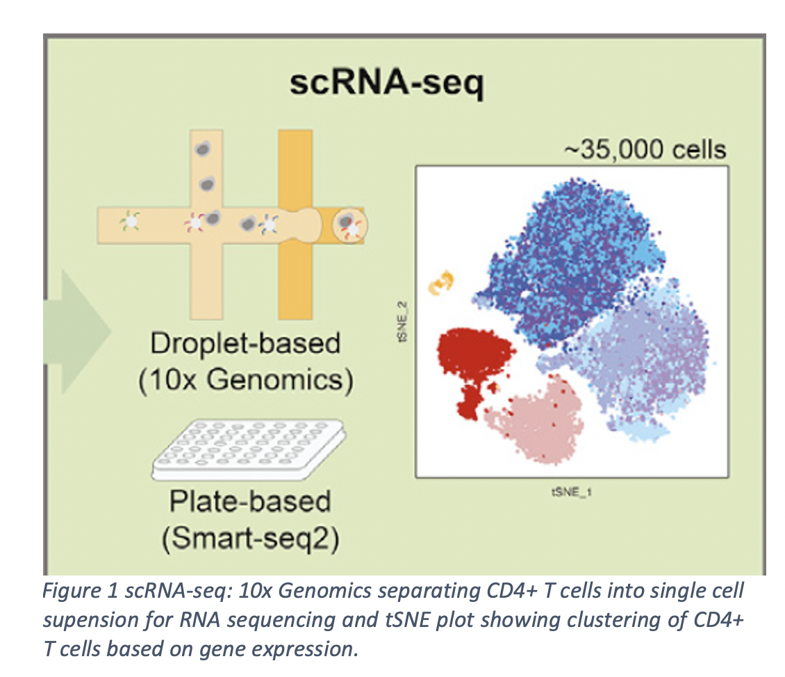
This technique measured all the genes expressed by each individual T cells. Based on the genes each T cell expressed, they were able to group cells expressing similar genes into clusters of either Treg cells or memory T cells (Tmem – antigen experienced T cells) as shown in the Figure 1. Further examination of these cell clusters revealed high diversity of Treg cell populations within the NLT and LT.
Pseudotime ordering analysis, an analysis that estimates the distance between cell transition states, was used to determine the trajectory of the Treg cells. Using this analysis ,transcriptomic adaptations occurring in Treg cells during their transition from the lymph node to non-lymphoid tissues could identify Treg cell subpopulations aligning in a continuous trajectory as shown in figure 2 and diverging towards their specific tissue fates carrying their respective address book e.g branchial lymph node (bLN) Treg (Lef1, Tcf7, Sell), mesenteric lymph node (mLN) Treg (Nfil3, Ccr8, Cxcr6, Gzmb), skin Treg (Sell, Tcf7, Rora and Tnfrsf9), and colon Treg (Sell, Tcf7, Rora and Tnfrsf9). These finding provide an easy tool to determine the fate of Treg cells in circulation and also demonstrated that the adaptation of Treg cells migrating to skin or colon depend on a shared transcriptional trajectory.
Using a melanoma mouse model, they could also show that, the core identity of NLT Treg cells is conserved between mouse and human. This genomic approach reveals a dynamic adaptation of T cells as they traffic across tissues and provide an open resource for investigating in vivo CD4+ T cell phenotypes in mouse and human, to ultimately harness NLT CD4 T cells as future therapeutic target.

REFERENCE
Miragaia, R. J. et al. (2019) ‘Single-Cell Transcriptomic of Regulatory T Cells Reveals Trajectories of Tissue Adaptation Resource Single-Cell’, pp. 493–504. doi: 10.1016/j.immuni.2019.01.001

Leave a comment