By Buntu Mlonyeni
Imagine trying to spot a grey cat in a foggy alley… This is how challenging it gets for doctors when trying to identify brain tumour MRIs. This time though, a human life is at risk. Brain tumours aren’t uniform masses , they are complex with distinct subregions. These subregions can either be an active tumour or edema. In MRIs, these subregions often share similar pixel intensities (shades of gray). So, you can imagine trying to differentiate between the subregions under intense pressure. Failure to correctly identify these regions can lead to surgical risks.
Traditional Ai tools try to segment these regions directly from raw MRIs but tend to struggle with this “fog” like how you would struggle identifying the cat. This leads to incomplete diagnosis.
To essentially “de-fog” MRI scans before analysis, researchers proposed the use of a conditional Generative Adversarial Network (cGAN). cGAN is a type of Generative Adversarial Network (Artificial Intelligence) where a generator and a discriminator both receive extra information (e.g. labels or images) as input. This allows the model to generate data conditioned on that input. Example: generating a specific digit or turning sketches into photos. The team built two cGANS,
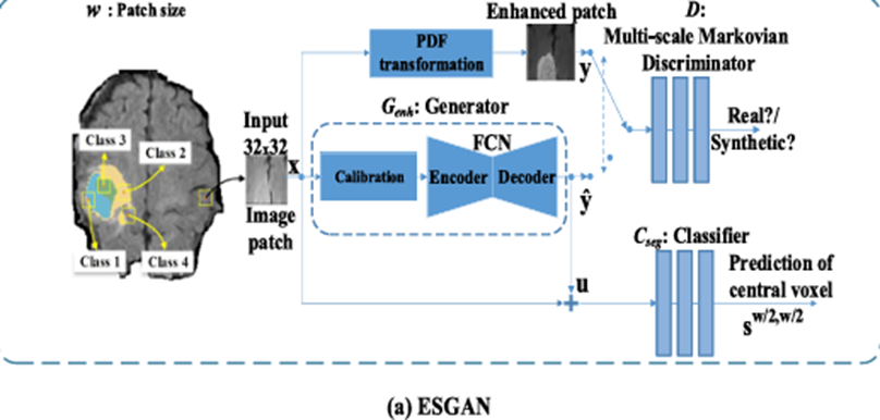
a) Enhancement and Segmentation GAN (ESGAN) – Tries to both improve the image and segment the tumour by learning from how patches (small image section of the tumour) are labelled
b) Enhancement GAN (EnhGAN) – Focuses on making a clearer higher-contrast version of the MRI image that makes it easier to see different parts of the tumour. It blends this new image with the original to highlight important areas and downplay less relevant ones.
The researchers used this model to identify the brain tumour (specifically Glioma) hidden in the gray shades.

The model tested on public datasets of brain MRIs with tumours worked (Glioma) well or better than existing methods (at least at the time this research was made) for segmentation. This to me matters beyond the lab. It isn’t about better Ai models or algorithms but about democratizing precision medicine. It reduces reliance on expensive multi-sequence MRIs, compensating for missing scans in low-resource clinics and provides a clearer visual for human-Ai collaboration. MRI scans usually cost a lot of money to a lot of people and this research tackles the silent issue in medicine of paying thousands of rands just to get a “maybe there’s something there”. The model working better than existing methods and models can improve and make diagnosis fast to prevent these issues
Reference
Hamghalam, M. and Simpson, A.L., 2024. Medical image synthesis via conditional GANs: Application to segmenting brain tumours. Computers in Biology and Medicine, 170, p.107982.


Leave a comment